Coloring Internal Cavities for Virtual Endoscopy
Omer Shibolet (1,2) and Daniel Cohen-Or (1)
(1) School of Mathematical Sciences, Tel-Aviv University, Ramat-Aviv 69978, Israel
(2) Algotec Systems Ltd, 4 Ha-Melacha St., POB 2408, Raanana 43000, Israel
Abstract
This paper describes a method for coloring voxel-based models.
The method generalizes the two-part texture mapping technique to
color complex and non-convex objects in a more natural way.
The method was developed for coloring internal cavities for
the application of virtual endoscopy, where the surfaces are shaped like a
twisted general cylinder in the macro-level, but with some folds and
bumps in the more detailed levels. Given a flat texture, the coloring method
defines a mapping between the 3D surface and the texture which reflects
the tensions of the points on the surface.
The core of the method is a technique for mapping a non-convex
surface to a convex one.
The new technique is based on a discrete dilation process that is fast
and robust, and bypasses many of the numerical problems common to
previous methods.
An updated version of the paper
-
Coloring Internal Cavities for Virtual Endoscopy(gzipped ps)
An mpeg movie of a Virtual Endoscopy simulation.

click here
Results
The figures below show a sequence of images of a sphere "growing" out
of a plan. Using our coloring technique, the texture seems to grow
together with the surface as if it a natural process. Note that we
do not apply a physical simulation but rather directly color the final
shape. The last image shows the same object textured by a naive two-part
mapping technique.
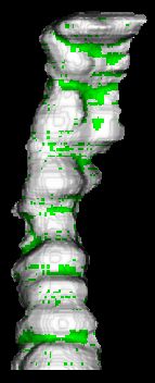
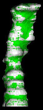
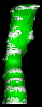
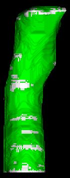
The following sequence shows a sample of the dilation process.
The original non-convex object slowly transforms into a convex shape.
The new voxels are colored in green.
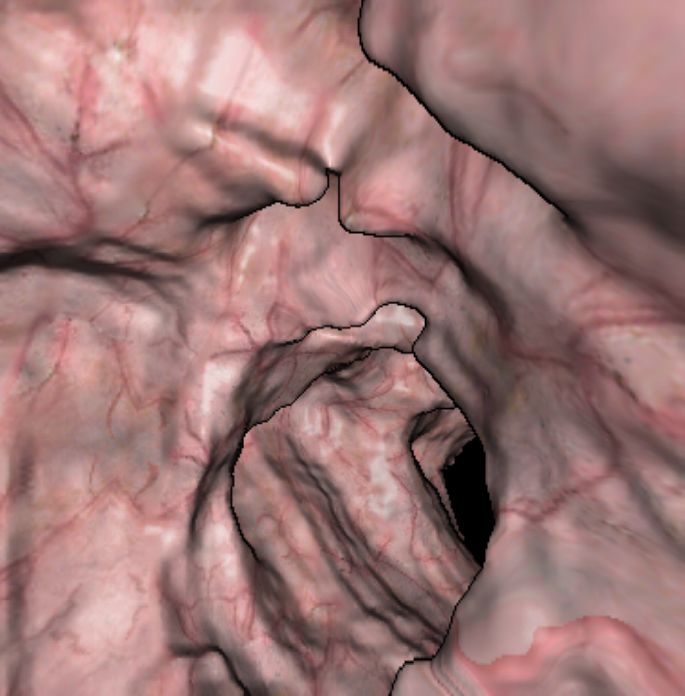
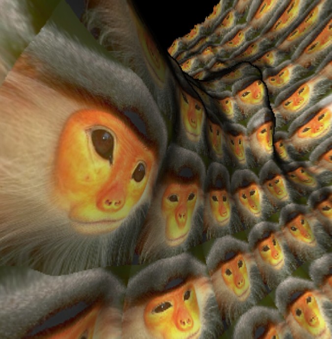
A high resolution image taken during interactive
navigation inside a region of the large intestine. This region of the
colon is colored using a replicated photograph taken during real colonoscopy
(endocopy of the colon).
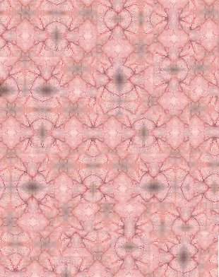
The replicated texture used to color the above image is
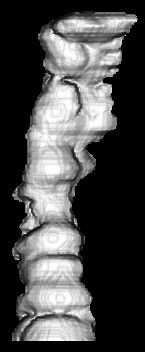
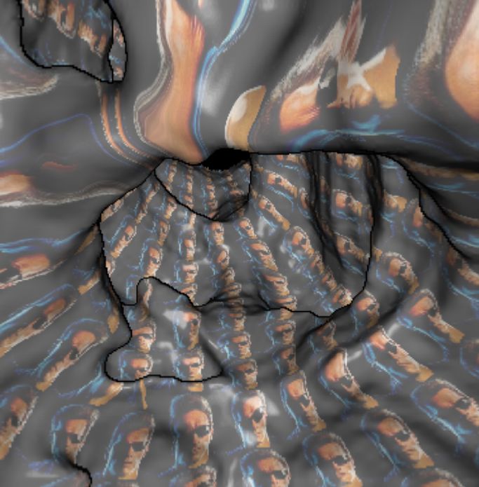
Another view of the colon, taken during virtual endocopy,
colored using a different texture.
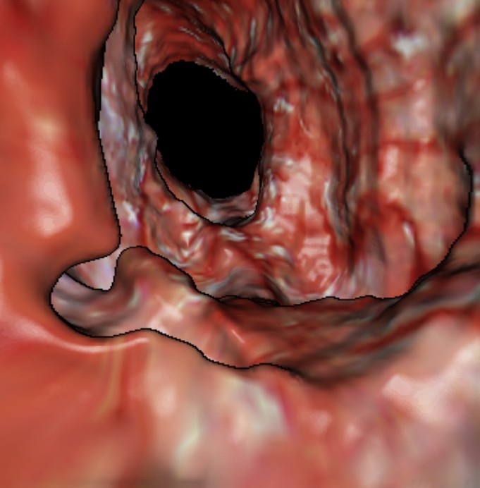
The following are colon regions colored with replicated images. Notice the
preservation of fine details in the process of coloring and rendering.
Address: Dept. of Computer
Science, School of Mathematical Sciences,
Tel-Aviv University,
Tel-Aviv 69978, Israel.
Phone: +972-3-640-8828
Fax: +972-3-640-9357
E-mail: {daniel | shib}@math.tau.ac.il, omer@algotec.co.il